

What Is Hyperopia?
Hyperopia, commonly called farsightedness, is a refractive condition in which the eye has difficulty focusing light directly onto the retina. This can cause blurry vision, particularly when viewing nearby objects.
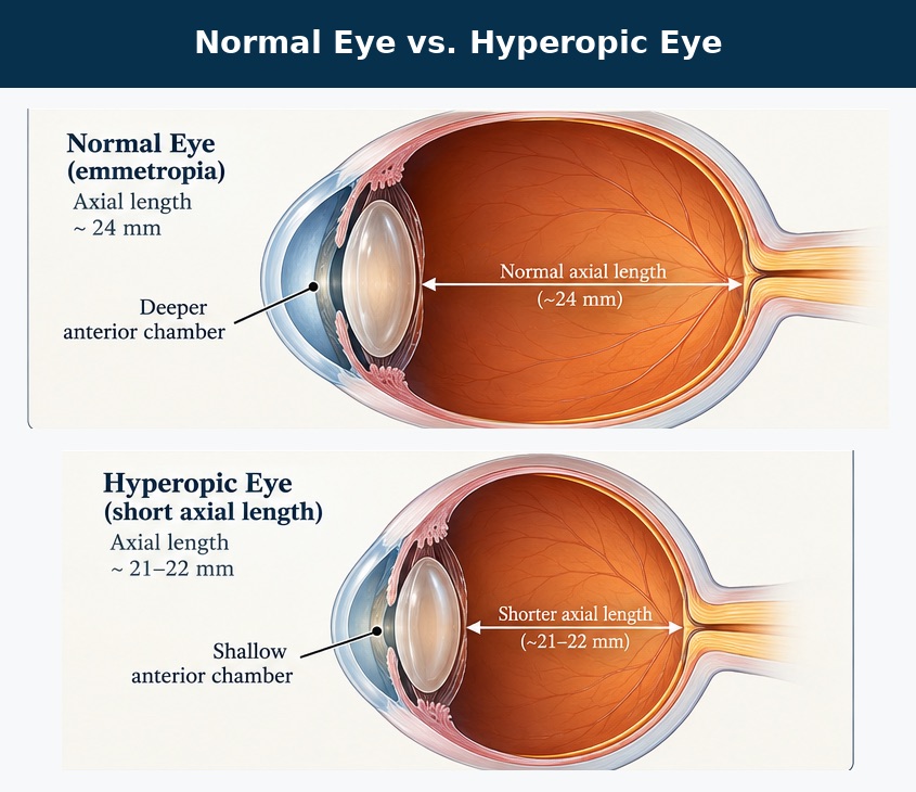
Hyperopia often occurs because the eye is shorter than average or because its optical focusing power is insufficient.
Although farsightedness is commonly treated with glasses or contact lenses, certain anatomical features associated with hyperopia can also increase the risk of a potentially serious eye condition known as angle-closure glaucoma.
How Does Farsightedness Increase Glaucoma Risk?
Unlike nearsightedness, which is primarily associated with open-angle glaucoma, farsightedness is associated with a greater likelihood of developing narrow drainage angles.
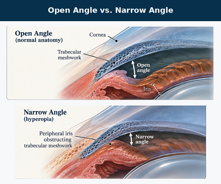
Inside the front of the eye, fluid called aqueous humor is continually produced and drained. Most of this fluid exits through a structure called the trabecular meshwork, located in the angle between the cornea and the iris.
In some farsighted eyes, the eyeball is relatively short, creating a more crowded arrangement of structures in the anterior chamber.
As a result:
- The anterior chamber may be shallower.
- The iris may sit closer to the drainage angle.
- The natural lens may contribute to crowding.
- The drainage angle can become partially or completely blocked.
When the iris obstructs aqueous drainage, pressure inside the eye may rise.
If this process causes optic nerve damage, the condition is called primary angle-closure glaucoma.
What Is the Difference Between Narrow Angles and Angle-Closure Glaucoma?
A common misconception is that having narrow angles means a person already has glaucoma.
In fact, these are different conditions.
Primary angle-closure suspect: The drainage angle is narrow enough to create potentially significant contact between the iris and drainage structures, but there is no evidence of pressure-related damage or glaucoma.
Primary angle closure: The angle is obstructed and there is evidence of elevated pressure or adhesions called peripheral anterior synechiae, but no glaucomatous optic nerve damage.
Primary angle-closure glaucoma: The angle-closure process has resulted in characteristic glaucomatous damage to the optic nerve.
Not everyone with narrow angles develops glaucoma. However, identifying individuals at risk provides an opportunity to prevent potentially serious complications.
Who Is at Greater Risk for Angle-Closure Glaucoma?
Although farsightedness is an important risk factor, it is not the only consideration.
Other recognized risk factors include:
- Older age.
- Female sex.
- Family history of angle-closure glaucoma.
- East Asian or Inuit ancestry.
- A shorter-than-average eye.
- A shallow anterior chamber.
- A relatively thick or forward-positioned natural lens.
The risk of developing angle closure generally increases with age because the natural lens gradually becomes thicker, potentially crowding the drainage angle further.
Importantly, not every farsighted patient has narrow angles, and angle closure can occur in individuals who are not farsighted.
What Are the Symptoms of Angle-Closure Glaucoma?
Angle closure may develop gradually and produce few or no noticeable symptoms.
This is known as chronic angle closure. Damage can develop over time without the patient realizing that anything is wrong.
However, some patients experience an acute angle-closure attack, in which eye pressure rises rapidly.
Symptoms may include:
- Sudden, severe eye pain.
- Blurred vision.
- Rainbow-colored halos around lights.
- Redness of the eye.
- Headache.
- Nausea or vomiting.
An acute angle-closure attack is a medical emergency. Patients experiencing these symptoms should seek immediate ophthalmic or emergency medical evaluation because untreated attacks can cause permanent vision loss.
How Are Narrow Angles Diagnosed?
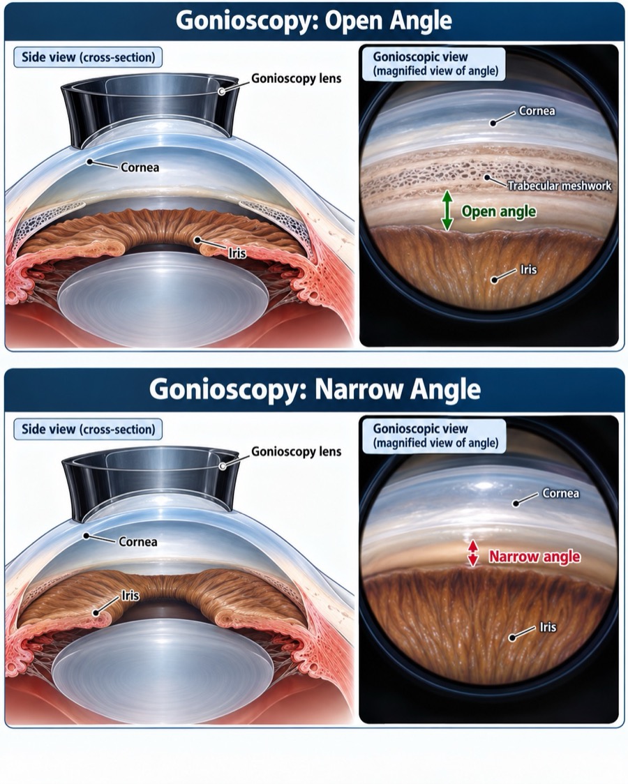
An important part of the evaluation is a procedure called gonioscopy.
During gonioscopy, an ophthalmologist places a specialized examination lens on the surface of the eye to directly examine the drainage angle.
This allows the physician to determine whether the angle is open, narrow, or closed.
Additional testing may include:
Anterior segment optical coherence tomography (AS-OCT): Produces detailed cross-sectional images of the structures at the front of the eye.
Ultrasound biomicroscopy (UBM): Provides additional information about deeper structures, including the ciliary body, when needed.
Eye pressure measurement: Helps assess whether obstruction of the drainage angle may be affecting pressure.
Optic nerve evaluation: Determines whether glaucomatous damage has already developed.
These findings help the ophthalmologist determine whether observation or treatment is appropriate.
Can Angle-Closure Glaucoma Be Prevented?
In many patients, the risk of angle closure can be reduced with appropriate treatment.
Two important approaches are laser peripheral iridotomy and lens extraction.
Laser Peripheral Iridotomy
Laser peripheral iridotomy, often abbreviated LPI, is a procedure that creates a small opening in the peripheral iris.
This opening helps equalize pressure between the front and back of the iris, reducing the pupillary-block mechanism that contributes to many cases of primary angle closure.
The procedure is generally performed in an outpatient setting.
However, LPI does not eliminate every mechanism of angle closure, and not all patients with narrow angles require preventive laser treatment.
For lower-risk patients, monitoring may be a reasonable approach following a comprehensive examination.
Cataract Surgery and Lens Extraction
The natural lens contributes significantly to anterior chamber crowding in many patients.
Removing the natural lens and replacing it with a thinner artificial lens can deepen the anterior chamber and widen the drainage angle.
For appropriately selected patients, particularly those with established angle-closure glaucoma or significant lens-related crowding, lens extraction may be an important treatment option.
Clinical practice guidelines recognize lens extraction as an effective treatment for selected patients with primary angle-closure disease.
Does Wearing Glasses Reduce the Risk?
Wearing glasses corrects farsightedness, but it does not change the underlying anatomy responsible for a narrow drainage angle.
A person can have excellent vision with glasses and still have narrow angles.
Similarly, having only a mild glasses prescription does not guarantee that the drainage angles are open.
An ophthalmologist must evaluate the actual anatomy of the anterior chamber to assess risk.
When Should Farsighted Patients Have Their Eyes Checked?
Patients who are farsighted should receive comprehensive eye examinations as recommended by their ophthalmologist.
Evaluation is particularly important for individuals with a family history of angle-closure glaucoma, increasing age, or previously identified narrow angles.
If narrow angles are detected, the ophthalmologist can recommend an individualized monitoring or treatment strategy based on the patient's risk factors and examination findings.
Protecting One’s Vision
Farsightedness is more than a glasses prescription. For some individuals, the underlying eye anatomy may increase susceptibility to angle-closure glaucoma.
Fortunately, narrow angles can often be recognized before permanent damage occurs.
At Inland Glaucoma Center, patients can receive detailed glaucoma evaluations to assess the drainage angle, optic nerve, and overall risk of disease.
If one has been told that their eyes have narrow angles, or if they are farsighted and concerned about glaucoma, schedule a comprehensive evaluation to discuss more with our ophthalmologists.
Works Cited
- American Academy of Ophthalmology. Primary Angle-Closure Disease Preferred Practice Pattern. 2026. Read guidelines.
- American Academy of Ophthalmology EyeWiki. Primary vs. Secondary Angle Closure Glaucoma. Updated May 18, 2026. Read article.
- American Academy of Ophthalmology. Primary Angle-Closure Disease Preferred Practice Pattern. Ophthalmology. 2021. View publication.
- American Academy of Ophthalmology. Laser Peripheral Iridotomy in Primary Angle Closure. Ophthalmology. 2018. View evidence review.
- American Academy of Ophthalmology. What Is Glaucoma? Patient education.