Open Angle Glaucoma
Open-angle glaucoma is the most prevalent type of glaucoma and a major contributor to irreversible blindness globally. This condition develops gradually, often without noticeable symptoms until significant vision loss occurs. It is primarily caused by damage to the optic nerve, typically resulting from elevated intraocular pressure (IOP) due to impaired drainage of aqueous fluid within the eye. Effective eye pressure management is crucial in mitigating the risks associated with this disease.
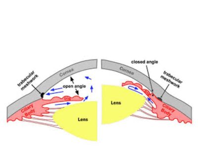
In a healthy eye, aqueous fluid flows continuously through the trabecular meshwork—a microscopic drainage channel located in the angle between the iris and the cornea. In cases of open-angle glaucoma, this drainage angle remains 'open,' yet the trabecular meshwork becomes partially obstructed, leading to a slow increase in eye pressure and progressive optic nerve damage. Understanding glaucoma treatment options is essential for preserving vision and managing this condition effectively.
Types of Open-Angle Glaucoma
1. Primary Open-Angle Glaucoma (POAG)
This is the classic and most common form of glaucoma known as open-angle glaucoma. Over time, the drainage system of the eye becomes less efficient, leading to increased eye pressure and slowly damaging the optic nerve. POAG often develops painlessly and without vision changes until advanced stages, highlighting the importance of effective eye pressure management and timely glaucoma treatment.
2. Normal-Tension Glaucoma (NTG)
In this type of glaucoma, optic nerve damage can occur even when eye pressure remains within the normal range. Factors such as reduced blood flow to the optic nerve, vascular dysregulation, or genetic predisposition may contribute to the condition, highlighting the importance of eye pressure management in effective glaucoma treatment, particularly for open-angle glaucoma.
3. Secondary Open-Angle Glaucoma
This occurs as a result of another eye condition or external factor—such as inflammation, steroid use, trauma, or pigment dispersion—that obstructs fluid drainage and raises eye pressure, which is crucial for effective eye pressure management. Understanding these factors is important in the context of glaucoma treatment, particularly for those with open-angle glaucoma.
Diagnosis
Because open-angle glaucoma progresses silently, regular comprehensive eye exams are critical for early detection and effective eye pressure management. Diagnosis typically involves: Measurement of intraocular pressure (tonometry), examination of the optic nerve (ophthalmoscopy or OCT imaging), visual field testing to detect early vision loss, corneal thickness measurement (pachymetry), and gonioscopy to assess the drainage angle. These steps are essential for appropriate glaucoma treatment.
Treatment Options
Although glaucoma damage cannot be reversed, early and consistent glaucoma treatment can slow or prevent further vision loss. This treatment primarily focuses on eye pressure management to protect the optic nerve, especially in cases of open-angle glaucoma.
1. Laser Therapy
Selective Laser Trabeculoplasty (SLT) is a highly effective and minimally invasive laser treatment used in glaucoma treatment, particularly for managing eye pressure in patients with open-angle glaucoma. This procedure improves fluid outflow through the trabecular meshwork and is often recommended as a first-line or adjunct therapy, potentially reducing the need for daily drops.
2. Eye Drops
Medicated drops are typically the first line of glaucoma treatment, particularly for managing eye pressure in conditions like open-angle glaucoma. These treatments function by either reducing fluid production or enhancing drainage. Common types include prostaglandin analogs, beta blockers, alpha agonists, and carbonic anhydrase inhibitors.
3. Oral Medications
Oral medications for glaucoma treatment are typically utilized when eye drops and laser therapies fail to adequately control intraocular pressure (IOP). These systemic drugs play a crucial role in eye pressure management by reducing the production of aqueous humor, the fluid found inside the eye.
Carbonic Anhydrase Inhibitors (CAIs) are the primary oral drugs employed for managing glaucoma, particularly in cases of open-angle glaucoma. They work by inhibiting the enzyme carbonic anhydrase in the ciliary body, which subsequently decreases the production of aqueous humor.
Common oral CAIs include:
Acetazolamide (Diamox) – The most widely used oral medication for glaucoma, available in both regular and extended-release forms.
Methazolamide (Neptazane) – This drug operates on a similar mechanism but is generally better tolerated, resulting in fewer side effects.
4. Minimally Invasive Glaucoma Surgery (MIGS)
MIGS procedures—such as the iStent, Hydrus Microstent, or OMNI canaloplasty—are effective for eye pressure management and enhance natural drainage pathways with minimal tissue disruption. These glaucoma treatment options are often performed during cataract surgery, providing added benefits for patients with open-angle glaucoma.
5. Conventional Glaucoma Surgery
In advanced or unresponsive cases of open-angle glaucoma, traditional procedures such as trabeculectomy or glaucoma drainage implants (Ahmed or Baerveldt valves) create new drainage pathways for effective eye pressure management.